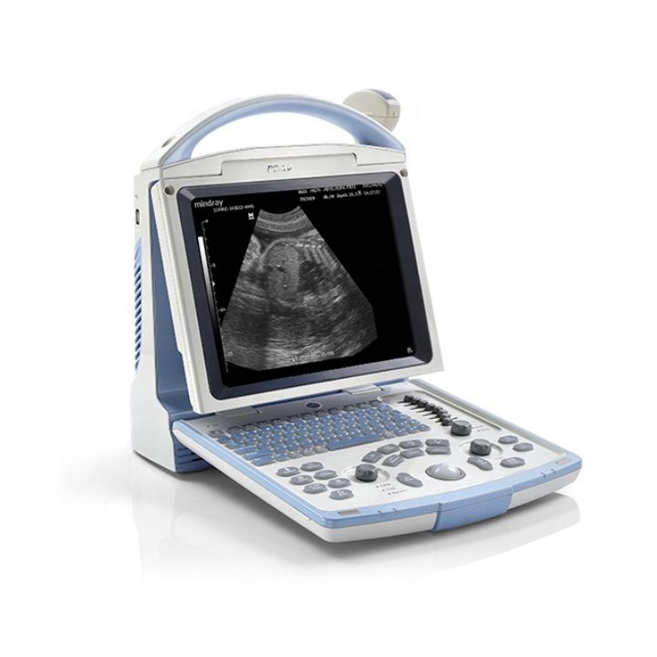
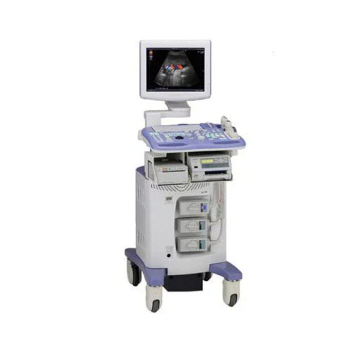
Mindray Dp 10 Portable Ultrasound
MINDRAY DP-10 B/W system with 1-probe connector, 12.1″ high resolution LED monitor, 2 USB port, 1 VGA OUT port, 1 video OUT, 1 S-Video OUT, software, without probe DP-10 slim and smart Good image quality, easy to use and lightweight to take it anywhere. It combines fashionable design, diagnostic confidence and economy.
Display modes and imaging processing:
Broadband, multi-frequency imaging: B, B/B, 4B, B/M, M
Imaging processing:
- multi-frequency probes for 2D imaging modes
- iClear: speckle reduction image
- THI (Tissue Harmonic Imaging) with convex probe 33976 only
- TSI (Tissue Specific Imaging)
- zoom function: spot zoom, pan zoom
- ExFOV (Extended field of view) Extended view of the anatomical structure on all convex and linear probes allows to discover better diagnostic information.
- iTouch (Auto image optimization)
- colour map
User interface
- alphanumeric QWERTY keyboard with backlight
- trackball: speed presettable
- 8-segment TGC
- user-defined blank keys: shortcut for easy access to menus
iStation™ intelligent workflow
- Intelligent patient data management system
- integrated search engine for patient data
- detailed patient information review
- intelligent data backup/restore
- patient data/image sending, data deleting (US B , DVD, DI COM)
- exam managing: activate exam
Storage/review mode:
- image archive on hard disk and USB mobile storage medium, temporary saving in cine memory, directly transfer report to PC
- 2 USB ports
- multiple image formats: BMP, JPG, DCM, FRM, AVI, DCM, CIN
- iVision: demo player
- Cine review: auto, manual (auto review segment can be set), supports linked cine review for 2D, M images
- Cine memory capacity (max.)
- clip length presettable: 1-60 s
- B mode: 11959 frame
- M mode: 110.0 s
- Supplied with GB, IT manual and CD manual (GB, FR, IT, ES, PT, DE, RU, BR).
iScanHelper function:
- Dedicated inbuilt tutorial function to enhance ultrasound experience.
- wide selection of application-specific exam planes
- anatomical illustrations
- standard ultrasound images
- scanning reference pictures
- tips on scanning and diagnosing

